Upregulation of Psoriasinin Cholesteatoma associated with inflammation, bone destruction and severity of disease: Histo-cyto-chemical and ultrastructural studies
DOI:
https://doi.org/10.56042/ijbb.v60i8.4299Keywords:
Bacterial localization, Cholesteatoma, Histo-cyto-chemistry, Psoriasin, Tympanomastoidectomy, UltrastructureAbstract
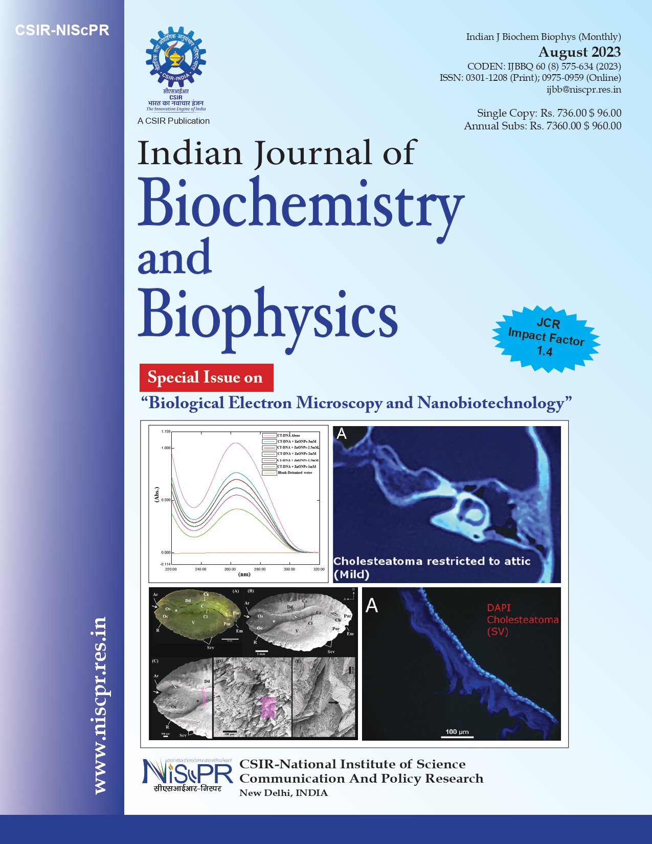
Cholesteatoma remains a mystery and the factors triggering and propagating it unknown. This study was aimed to evaluate the role of Psoriasin and pro-inflammatory cytokines in progression of middle ear cholesteatoma. In this study 18 patients (12 cases of cholesteatoma and six cases of posterosuperior retraction pocket (PSRP) without cholesteatoma) were included in the study. Clinical aggressiveness was evaluated on-table under operative microscope by evaluating erosion of ossicles, invasion of surrounding structures. The classification of disease was also based on the radiological (CT-scan) findings. Based on clinical aggressiveness, the cholesteatoma cases were divided into mild, moderate, and severe. The tissues were processed for histopathological, ultrastructural and immunohistochemical analysis. The Psoriasin expression in cholesteatoma tissues was significantly higher (P< 0.05) compared to the PSRP tissue and increased with the increasing severity of the disease. The pro-inflammatory cytokines (IL-6 and IL-1β) were also upregulated along with vascular endothelial growth factor and Matrix metalloproteinase-13 in cholesteatoma cases compared to control. Transmission Electron Microscopic images of ultra-thin sections of the tissues showed numerous secretory vesicles, most likely containing cytokines and a unique ultrastructural feature of bacterial localization within the membrane pocket was seen, which gave direct evidence of bacterial infection in the middle ear cholesteatoma.